The First Fully Integrated Single-Cell Spatial Solution
Key Applications

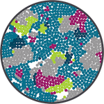
Cell Atlasing/Cell Typing
Discover and map cell types using expression profiles of known RNA and protein targets

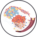
Disease State
Visualize and quantify molecular (RNA / protein) and cellular organizational changes in a tissue

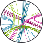
Ligand-Receptor Interaction
Analyze expression and interactions of up to 100 classic ligand-receptor pairs between interacting cells

Biomarker Discovery
Reveal differential gene expression and pathways in the same cell types depending on their spatial location

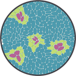
Tissue Microenvironment
Understand cellular neighborhoods by examining individual cells and their interacting neighboring cells
How It Works

Going from tissue sample to single-cell and subcellular insights has never been easier. With CosMx SMI, researchers can choose between preparing fresh frozen (FF) or even formalin-fixed paraffin embedded (FFPE) tissue samples with our 1000 plex RNA assay or 64-plex protein assay.
This streamlined workflow integrates with standard ISH protocols and eliminates the need for tissue expansion or clearing, cDNA synthesis, and amplification. Once tissue samples have been prepared, researchers can load their slides and associated CosMx reagents and allow the platform to perform the automated cyclic in situ hybridization chemistry.
While the tissues are being imaged on the CosMx SMI, a machine learning-driven algorithm performs cell segmentation on the images in parallel. Once a CosMx run has completed, these image files are ready to be analyzed and interpreted on AtoMx SIP with a few clicks.
Project Support
For questions and to discuss new projects please contact us via our group email: CCRSPITR@mail.nih.gov
Contact SpITR